By James Van Howe
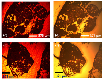 Left: Reflection images of a histopathology slide corresponding to skin tissue using a low-cost, portable, lens-free off-axis holographic microscope. Right: Conventional reflection-mode microscope image of the same specimen using a 4X objective lens (NA: 0.1). Image from Biomedical Optics Express.
This post originally appeared on Jim’s CLEO Blog and is reproduced with permission from the author.
Research performed in the Ozcan group at UCLA holds a unique place in the field of optics and photonics. Besides the typical pursuit of advancing optical technology, another major initiative of this photonics group is solving problems of global world health, particularly in resource-poor countries.
Early September marked a milestone for the UCLA group as they published work on a compact, low-cost (~$100 USD of parts), dual-mode microscope with 2 micron resolution in Biomedical Optics Express (also written up in a recent OSA press release). The key to making such a low-footprint, low-cost, lab-grade device is using holographic microscopy. The image information stored in a hologram (the interference of the reflected or transmitted light from the specimen with a reference beam) requires no lenses, drastically reducing the weight, size, and overall expense of the device. A computer reconstructs the wavefront reflecting from (or transmitting through) the sample instead of a lens (see fig below). The impact to world health will be increased blood-diagnostics, water quality tests, tissue screening and analysis, and other imaging diagnostics in areas where microscopes currently are not available due to cost and/or remoteness of location. Getting more microscopes into the hands of health workers may have large impacts for heading off disease outbreaks as well as treatments for individuals.
The idea of using holograms in microscopy is not new. In fact it was the quest for higher resolution in electron microscopy which prompted Dennis Gabor to devise wavefront reconstruction by holography in 1948. Gabor coined the word “hologram” which translates “whole message” to emphasize the amount of information that is stored in this very special interference pattern. For a brief history of holography from its roots in microscopy, its development through radar, and its boom in mainstream art and media in the 60′s and 70′s , see Jeff Hecht’s 2010 OPN article.
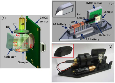 Schematic of the 200 gram microscope developed by the Ozcan group in reflection mode. LD: laser diode, PH: pin hole, BC: Beamsplitting Cube. Note the two AA batteries as the power source as well as for scale. Image from M. Lee, O. Yaglidere, and A. Ozcan, Biomedical Optics Express, 2, 2721 (2011).
What makes the Ozcan group’s work so special is not the use of a fundamentally new technique, but clever and impressive engineering. This holographic microscope is small, inexpensive, and can work in both transmission and reflection mode. The transmission mode of the current device is similar to an earlier work by the Ozcan group- a cell-phone microscope. In the summer of 2010, the UCLA group published work in Lab on a Chip demonstrating a clever attachment to an ordinary cell-phone which could convert it into lab-grade microscope (see the youtube short below). By employing digital holographic microscopy, the group was able to produce a 38 gram attachment without any lenses, lasers, or bulky optics, which when incorporated with the cell phone camera, produced hologram on the cell phone detector array. The idea is that the hologram data would be sent over the same cell phone to the closest hospital/analysis station, a computer would process the hologram to extract the image information, and then the image would be sent back to the same phone, all within seconds of placing the sample to be analyzed into the device.
Though the current device cannot be so easily integrated onto a phone, the additional benefit of reflection-mode operation makes up for its “bulkiness.” By operating in reflection-mode, the new microscope is additionally suited for imaging optically dense media like tissue, something not possible using in-line transmission holography due to spatial distortions in the reference wave… To read the full original post, click here.
Posted: 3 October 2011 by
James Van Howe
| with 0 comments