By Frank Kuo
 This post originally appeared on CLEO BLOG by Frank Kuo and is reproduced with permission from its author.
To probe new scientific frontiers, we need new technology. On the other hand, the advance of the technology relies on the solid scientific foundation. Countless examples have shown us that science and technology evolve together to give us wonders and a better understanding of the universe and nature. Looking back at 2012, similar stories happened in the laser and optics arena – Lasers are extending the working wavelengths into shorter (X-rays) and longer (THz) domain and probe new scientific frontiers. With this advance, we can have a better grasp of our nature.
Femtosecond X-ray free electron lasers, the most established source to generate “coherent (laser-like) X-ray”, relies on a gigantic synchrotron. In brief, a bunch of high-energy electrons from the synchrotron is sent into a long tunnel made of magnets. The tunnel, often more than 100 meters, is called undulator. The magnets are arranged in a way such that they create an alternate magnetic field to wiggle the electrons and force them into emitting X-rays. The wiggles are tuned to the wavelength of the X- ray and creating a feedback mechanism – this radiated X-ray acts on the electrons, concentrating them into smaller and tighter groups, and makes the electrons emit more X-ray coherently. Apparently, it is very similar to normal lasing scheme, in which the radiation in the cavity induces more radiations. The main difference is that in the case of X-ray, there is no cavity since no reflective mirrors are available in this wavelength region.
What excites us in 2012 is that this “new light” gives us a better way to elucidate the secret of our living nature. It is used to probe the structure of the proteins:
In the old days (well, even in nowadays), to peek at the precise structure of a protein with the atomic resolution, you need to grow a “sizable” protein crystal such that you can use crystallography to decipher its structure. The reason is straightforward – without a sizable crystal, it cannot withhold the constant bombard from incoherent X-ray interrogation. However, growing a crystal made of protein is not easy. People used to joke that if a graduate student grew a big crystal, then he/she bought his/her ticket to a Ph.D. degree. That sort of tells us the difficulty.
With the advance of the femtosecond X-ray FEL, it gives us a new window. We do not need monster size crystal anymore. This “new light” is coherent which means it is intense and produces high-quality diffraction. On the other hand, its pulse duration is short (within tens of femtoseconds) such that the entire process ends before the onset of substantial radiation damage.
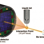
Researchers from SLAC National Accelerator Laboratory demonstrate this new technology beautifully to us. They use a 430 m long undulator to create femtosecond X-ray, either 5 fs or 40 fs pulse, with the wavelength of about 9.4-keV. The target is a liquid jet filled with micro-crystal of lysozyme (each crystal is less than 1 micrometer by 1 micrometer by 3 micrometers). The experimental setup is shown conceptually in figure 1. This result is very satisfactory and fits well with data that are gathered from traditional X-ray crystallography (figure 2).
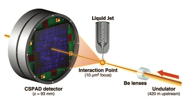 Figure 1. The experimental set up of new crystallography realized by femtosecond X-ray FEL. Courtesy of S. Boutet and et al. in Science 337 362 (2012).
 Figure 2. Part of the protein structure (electron density map) at 1.9 Angstrom resolution of lysozyme elucidated by this new technique. Courtesy of S. Boutet and et al. in Science 337 362 (2012).
Trivia about X-ray optics:
X-ray, although belongs to the family of electromagnetic radiation, has some interesting property which amuses me quite a lot.
-
First of all, it has index of refraction very close but smaller to 1. Figure 3 shows how index of refractions varies with the frequency of light. This means that its phase velocity is faster than light! Shocking! For people who are interested in this topic in more details, check this link out.
-
Normal mechanism we use to focus visible or IR light simply does not work in X-ray regime. For the reflective optics, X-ray only reflects efficiently if the incident angle is very shallow (close to 90 degree). If the incident angle is too small, the x-ray penetrates into the optics without being reflected. This phenomenon creates the need of special reflective optics, such as Kirkpatrick-Baez mirror to focus the X-ray (figure 4).
-
Just like I mentioned in above, the index of refraction of X-ray is just slightly below 1, we have to re-think how to focus X-ray if we want to use refraction principle. First of all, you have to think air as a glass, since air has index of refraction equals to 1, and the material has something smaller than one. A collimated X-ray will focus when propagating from the material to the air. Secondly, one refraction is hardly enough, since the index of refraction in the material is just slightly smaller than 1. As a result, to create strong optical power, you need many air bubbles in tandem buried in the material. The Be lens used in figure 1 has the structure like the picture shown in figure 5.For more descriptions on the X-ray lenses, check out this website: http://www.xradia.com/technology/basic-technology/focusing.php
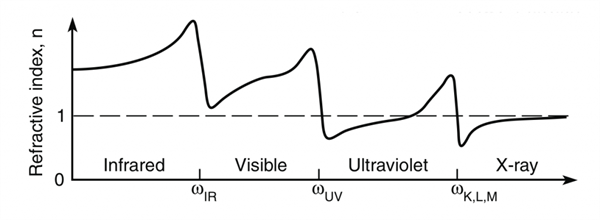 Figure 3. The index of refraction vs. wavelength. Courtesy of D. Attwood, an online resource.
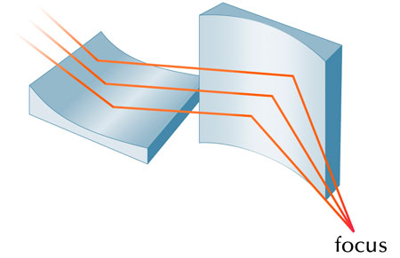 Figure 4. An Kirkpatrick-Baez mirror to focus the X-ray.
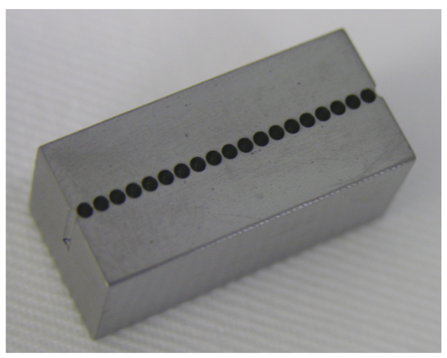 Figure 5. A beryllium lens with 20 lenslets in a 20 mm x 10 mm x 10 mm substrate (left). The lenslets are 5 mm deep. Courtesy of A. Khounsary and et al. at Beryllium and lithium X-ray lenses at the APS, an SPIE 2006 paper.
DISCLAIMER
The opinions expressed herein are those of the author and do not represent the Optical Society of America (OSA) or any OSA affiliate.
Posted: 16 February 2013 by
Frank Kuo
| with 0 comments